Previous
Next
1 of 3

Mars Bioimaging

Mars Bioimaging

Mars Bioimaging
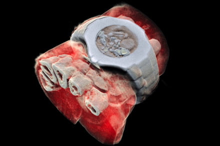
Your next X-ray could be a whole lot more colorful — and you’ve got Europe’s CERN physics lab and a New Zealand startup to thank for it.
The color X-ray technology, which could help improve the field of medical diagnostics, utilizes particle-tracking technology developed for CERN’s Large Hadron Collider. According to the European Organization for Nuclear Research, known as CERN, it could be utilized to produce clearer, more accurate images compared to the traditional black-and-white X-rays hospital doctors have been using routinely since at least the 1930s. This should prove particularly valuable in diagnosing diseases, including cancer and heart disease, because it provides greater detail concerning the body’s chemical components.
The technology is now being commercialized in the form of a dedicated scanner by a New Zealand startup called Mars Bioimaging, which recently performed the world’s first ever color X-ray of human body parts — in this case, the ankle and wrist.
“Mars scanners use a detector that uses the color or energy information of the x-rays that traditional x-ray detectors don’t use,” Professor Phil Butler, a physicist working at New Zealand’s University of Canterbury who helped invent the Mars scanner, told Digital Trends. “This color or energy information from the x-ray, also known as spectral information, is used to distinguish different atoms or materials from each other, [such as] calcium from iodine. In addition, Mars scanners have a much smaller pixel size, meaning its possible to generate 1000x more information than existing CT systems for the same dose.”
Phil Butler co-developed the scanner with his son Anthony Butler, a radiologist and professor at the Universities of Canterbury and Otago. The technology is already available in the form of a small-bore scanner for carrying out medical research. The pair now plan to produce commercially available body part scanners for use on patients.
“There are wide applications for this technology,” Phil Butler continued. “We have already demonstrated using our small-bore system that it can be used for investigating composition of plaques that cause strokes, measuring the deterioration of cartilage, viewing bone-metal implant interfaces, and detecting small cancers in mice.”
Hopefully it won’t be long before this is a standard piece of equipment in hospitals everywhere.
Editors’ Recommendations
- X-ray laser heats water to 180,000 degrees in a fraction of a second
- The best Dolby Atmos movies, from ‘Mad Max: Fury Road’ to ‘Gravity’
- Fujifilm X-H1 review
- Win an iPhone X and rep your soccer team with Speck Presidio World Grip cases
- MIT can charge implants with external wireless power from 125 feet away

